When Only AI Can See the Cancer
Vivian Manning-Schaffel New York Magazine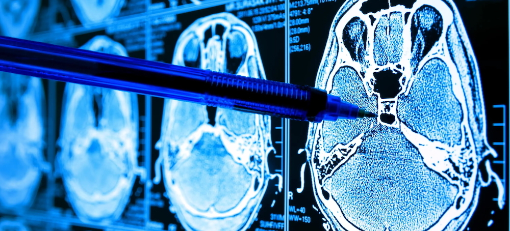 "As artificial intelligence continues to evolve its impact on cancer is likely to grow exponentially." (photo: Parkridge Health)
The cancer had been missed on at least five imaging tests over ten months — an all-too-common occurrence because it is exceedingly difficult to visualize pancreatic cancer with current screening technology. About 40 percent of pancreatic tumors less than two centimeters in size are missed on CT scans, according to a 2021 study published in European Radiology. When patients turn up at the hospital suffering from a constellation of symptoms that could be signs of many diagnoses, it’s not unusual for busy ER radiologists to miss pancreatic abnormalities. In 2022, Michael B. Wallace, an interventional endoscopist from the Mayo Clinic in Jacksonville, Florida, conducted a study to determine just how often pancreatic cancer could be missed or go unrecognized when doctors order imaging to look for more likely diagnoses, such as kidney stones, appendicitis, and gallbladder infections. In examining CT scans and MRIs from one month to three years before pancreatic-cancer patients were diagnosed, Wallace discovered that as many as half of the scans indicated an abnormality in the pancreas that could have prompted more testing.
After hearing her diagnosis, Jackson says, her gastroenterologist could barely look her in the eye: “He was so heartbroken. He was like, ‘I must’ve missed something. How could this happen?’”
These kinds of misses are a major reason pancreatic cancer is projected to be the second deadliest in the United States by 2030. Only 12 to 15 percent of patients survive it because it is too often found too late. Approximately 67,530 people will be diagnosed with pancreatic cancer this year, with Black Americans, like Jackson, more likely to develop pancreatic cancer than any other group, according to the National Cancer Institute SEER data.
But recently, those who study the disease have found themselves cautiously optimistic. Part of this is due to the FDA’s approval of a highly effective daily pill (daraxonrasib) for metastatic pancreatic cancer, an event that grabbed headlines over the summer. But the positivity also stems from less publicized but still significant advances in early pancreatic-cancer detection. Over the past few years, researchers have been harnessing AI to develop new screenings that flag concerning or cancerous lesions on scans before they’re visible to the human eye. There is already promising data on this technology, and experts say the long-term results have the potential to be transformative.
Ajit Goenka, M.D., a specialist in nuclear medicine and abdominal imaging at the Mayo Clinic in Minnesota, has had to break pancreatic-cancer diagnoses to scores of patients who had previously received seemingly normal results on abdominal imaging — an experience that always leaves him both frustrated and ashamed on behalf of his profession. Every day, his team notices subtle signs of pancreatic cancer on imaging that should have aroused suspicion months, if not years, before. “The first time 80 to 85 percent of people hear pancreas cancer, they have unfortunately already lost the battle,” he says. “We need to be able to find it at a stage where we can flip that equation from 85 percent of patients presenting too late, to about 85 percent presenting at a stage where a curative treatment, like surgery, could be a part of the equation.”
Knowing pancreatic cancer can lie in wait on a cellular level for years before becoming symptomatic or detectable on imaging, Goenka set out to determine whether AI could sift through CT images to detect easy-to-miss signs of cancerous tumors, neoplasms (abnormal tissue growths), or precancerous cells. “AI is very good at looking at grains of sand, whereas we as human beings are better at looking at the beach,” Goenka explains.
He built an AI framework called REDMOD, which stands for radiomics-based early-detection model, training it with both CT scans taken from patients who were diagnosed with pancreatic cancer between three and 36 months after their scans and with control scans from healthy patients spanning approximately 15 years of clinical practice at the Mayo Clinic. By analyzing subtle patterns in the brightness and texture of the pancreas that are difficult for the human eye to appreciate, REDMOD can spot and flag subtle signs of cancer roughly 16 months earlier and at nearly double the rate of specialist radiologists. “You could ask ChatGPT to, in one minute, read a thousand-page book and find the page that says ‘red bird,’ but if I ask you, a human, to read a thousand-page book and, in retrospect, ask you to tell me the page that says ‘red bird,’ that’s hard to do,” says Wallace. It took months for imaging to verify Jackson’s metaphorical “red bird,” while solutions like Goenka’s REDMOD could have done it in minutes. If it had been available to her care team, she might still have her gallbladder.
Like Goenka, Neil Marya, M.D., has a memory bank full of frustrating and heartbreaking patient conversations. He is the co-director of the program in digital medicine at UMass Chan Medical School, and he says he struggled to tell patients that even though their biopsies came back negative, they still had a high chance of having cancer in their bile ducts because the biopsies are not sensitive enough to detect early disease. “It’s really awful,” he says, referring to how much anxiety this uncertainty sparks in his patients. Even worse, by the time his patients’ biopsies turned positive, it was often too late to remove the cancer or the organ.
Marya turned to AI to develop a way to accurately flag bile-duct lesions that are visibly suspicious or cancerous and are likely to come back with a negative biopsy. Using video data from previous endoscopies, Marya created an AI-based tool for cholangioscopies (the bile-duct equivalent of an endoscopy) that can analyze bile ducts for suspicious changes in real time. Along with his childhood friend Patrick Powers, a medical-application developer at UMass, Marya trained an AI tool (built using hardware from Nintendo Switch) to recognize various types of tumors and suspicious lesions he often spots during cholangioscopies by feeding it 20,000 random images from 130 procedures conducted at UMass and the Mayo Clinic. The fruit of his labor is SMART-AI, a plug-and-play hard drive that works with existing cholangioscope machines to visualize early cancerous growths better than biopsies do. A study Powers and Marya conducted in April 2026 suggested SMART-AI was remarkably effective; its first human trial was 88 percent accurate in diagnosing bile-duct cancer, compared with regular biopsies that were only 67 percent accurate.
For many cancers, preventive screening is a universal recommendation because current imaging is highly effective in catching disease early. The American Cancer Society recommends that women over age 45 with an average risk of breast cancer receive mammograms each year and that high-risk women over age 30 receive both an MRI and a mammogram. ACS also recommends colonoscopy screenings for anyone over age 45, and the Centers for Disease Control recommends that all former heavy smokers and people over 50 receive low-dose CT scans to screen for lung cancer. At this time, preventive screening for pancreatic cancer is incredibly targeted (reserved only for those who have relatives with a pancreatic-cancer diagnosis or who test positive for a high-risk gene mutation, or both). According to Dr. Vineet Syan Rolston and Dr. Mengmeng Du, both at Memorial Sloan Kettering Cancer Center, this is largely because pancreatic cancer is rare and screening wouldn’t necessarily find the cancer anyway. Currently, screening for high-risk patients at MSK includes blood tests and annual imaging that alternates between a magnetic-resonance cholangiopancreatography (radiation-free imaging that gives detailed images of the pancreatic and bile ducts) and an endoscopic ultrasound, in which a thin, flexible tube is passed through the mouth so a small ultrasound camera can look at the pancreas up close. But Wallace sees these new AI tools as having the potential to simplify this process and expand it to a wider population. “These AI systems are quite accurate,” says Wallace. “You can imagine putting this software in the emergency department and just running it on every scan being done and saying, ‘Flag it if it looks like there might be early pancreas cancer.’”
In China, Xiaomeng Li, Ph.D., has been using AI to help doctors more accurately spot rare cancers in biopsy tissue. Her software framework, called PRET, scans digital images of tissue samples and flags potential cancers or other abnormalities; it could help less experienced doctors, or those in remote or small hospitals, diagnose patients faster and more accurately. Careful to mention that PRET isn’t meant to replace specialist expertise or stage cancer, Li, of the Hong Kong University of Science and Technology, is nonetheless enthusiastic about its use as a validation or verification tool. Thus far, PRET has yielded impressive results: It has a 98 percent accuracy rate, outperforming 11 pathologists on lymph-node metastasis detection.
How long it may be until AI becomes a standardized element of screening for cancer — or for any medical problem — remains to be seen, but lately, progress to that end has been swift and significant. Between January and June of this year, the FDA approved 135 out of 181 AI-enabled radiology devices for medical purposes. And for the past few years, a radiology company called RadNet has been providing enhanced breast-cancer detection, an AI-powered mammogram, to clinics in New York and New Jersey. In 2025, to improve predictions of whether a woman will develop breast cancer over the next five years, the FDA approved Clairity Breast, an AI-powered platform that works with standard mammograms. The company received $43 million in funding to roll out the technology throughout the nation over the next few years so that leading imaging centers and health systems can have access to it.
Wallace says it took around three years for GI Genius, an AI-driven tool he helped develop to flag potentially cancerous polyps during colonoscopies, to go from an idea to FDA approval. He’s now at work on a large clinical trial of Cyst-X, an MRI-based AI algorithm that can ferret out pancreatic cysts — one of the few identifiable precursors to pancreatic cancer. Marya and Powers are still trying to clear various regulatory hurdles before a version of SMART-AI can be plugged into cholangioscope machines, and Li and her team have begun testing PRET at one hospital in mainland China and hope to expand to more hospitals soon.
As for Goenka, he’s in the midst of a large clinical trial of REDMOD that he hopes will help more of his patients be diagnosed with pancreatic cancer in time to benefit from surgery. “Early pancreatic cancer can leave the pancreas looking nearly normal,” says Goenka. “AI may be able to detect subtle patterns that humans cannot reliably see. In that sense, I do think AI can help scale human excellence.”